
MCATSS offers a wide range of solutions for macro-, micro- and nano-scale characterisation of surfaces, bulk materials and non-engineering related materials. We have access to high-resolution capabilities for quantitative and qualitative analysis.
microscopy
Optical Light Microscopy - ideal for broad, low magnification surface analysis of materials or for thin translucent materials.
Scanning Electron Microscopy (FE-SEM) - with EDX, WDS, BSE, ESEM, cryo-SEM, low vacuum mode - one of the most common methods in materials science, SEM is used for high magnification analysis of engineering and biological materials. Used in various modes, information gained includes details of microstructure, contamination, composition, failure mode, feature size, morphology etc.

Optical micrograph of etched steel. Image @50x magnification

FIB milled cross-section of a coating revealing sub-surface defects
Focused Ion Beam (FIB) - similar to SEM with electrons, FIB utilises a column of ions for a number of uses such assurface etching, TEM sample preparation and cross-section for sub-surface analysis.
Transmission Electron Microscopy (TEM) - is a technique used to image ultra thin specimens and is applicable to physical, chemical and biological applications. It is typically used to analyse microstructures, dislocations, atomic structures, tissue, pollen etc.
Micro-CT imaging - X-ray micro-CT uses x-rays to produce 3D cross-sections of an object without the need for destructive sample preparation. It is useful when investigating porosity, cracking or other features within the bulk of the material.
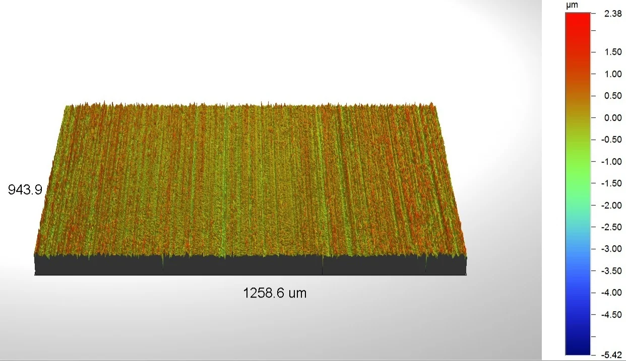
Surface profilometry analysis of a rolled steel plate using WLI
surface profilometry
Atomic Force Microscopy (AFM) - is an ultra-high resolution scanning probe method used to measure and image surface features in the sub-nm scale. Using various imaging modes, it can be used to analyse features such as grain structure, porosity, roughness, cross-linking, proteins, tissue, plants, conductive layers and semiconductors.
White Light Interferometry (WLI) and Confocal Microscopy - WLI and Confocal work in a similar way to produce 3-dimensional profiles with near-nm resolution. Useful information about a surface can be quickly obtained, including flatness, roughness, volume, peak/valley heights and skew. Complex profiles can often be imaged readily.
Please contact us if you have specific requirements that are not listed here.